
Analysis of Tumor Marker---Ferritin(FER)
Views:1309 Add time:2018-10-30 14:54:09
Ferritin(FER) is a globular protein complex of 450 kDa consisting of 24 protein subunits and is the primary intracellular iron-storage protein in both prokaryotes and eukaryotes, keeping iron in a soluble and non-toxic form [1]. It occurs normally in almost all tissues of the body but especially in hepatocytes and reticuloendothelial cells, where it serves as iron reserve. Ferritin is aslo present in the serum in minute amounts, where it appears to reflect iron stores in normal individuals.
A low serum ferritin value is thought to be the best laboratory indicator of iron depletion. Virtually all patients with low serum iron and low ferritin have iron deficiency. Serum ferritin is clincially useful in distinguishing between iron-deficiency anemia(serum ferritin diminished levels) and“anemia of chronic disease”(serum ferritin levels usually normal or elevated).[2,3] Serum ferritin is a good screening test in separating erythrocytes microcytosis due to iron deficiency(low values) from microcytosis related to thalassemia minor(normal or high values). An iron-depletion state with a decreased serum ferritin value is quite common among menstruating and reproductively active females and in children.
A high serum ferritin value is seen in hemochromatosis and other iron-overload states, as well as cute hepatitis, Gaucher disease, malignancies, and chronic inflammatory disorders.
Anti-Human Ferritin Monoclonal Antibodies
A new generation of unique anti-ferritin monoclonal antibodies, which was recently produced by CUSAg, makes possible the development of highly sensitive and rapid sandwich immunoassays. Our in-housechemiluminescent immunoassays have a linear detection range from 2.5 to 600 ng/mL. All recommended MAb combinations were evaluated in medium-scale clinical trials with blood specimens from anemia, normal and inflammatory patients.
|
Properties |
Specification |
|
Target species |
Human |
|
Host animal |
Mice Balb/c |
|
Cell line used for fusion |
Sp2/0 |
|
Immunogen |
Human Ferritin Protein |
|
Purification method, Purity |
Protein G affinity chromatography,>95%(SDS-PAGE) |
|
Presentation |
MAb solution in PBS (pH 7.4) |
|
Application |
CLIA, LETIA, LFIA etc. |
|
Catalog Number |
CSB-DA027BmN③, CSB-DA027BmN④ |
Calibration Curve
CLIA platform
All monoclonal antibodies were tested in pairs as capture and detection antibodies to select the best two-site MAb combinations for the development of a quantitative sandwich immunoassay. Calibration curves for several best two-site combinations are shown in Fig.1. Detection antibodies were labeled with horse radish peroxidase (HRP). The best selected MAb combinations for quantification of human ferritin are (capture-detection respectively):
MAb combination A: CSB-DA027BmN①- CSB-DA027BmN②;
MAb combination D: CSB-DA027BmN④- CSB-DA027BmN③.
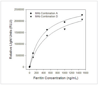
Fig.1 Calibration curves for ferritin in sandwich chemiluminescence immunoassay (CLIA)
Antigen: human ferritin
Capture MAbs-unlabeled.
LETIA platform
Anti-human ferritin monoclonal antibodieswere also evaluated by CUSAglatex-enhanced immunoturbidimetric assay (LETIA). A set of ferritin calibratorsreacts with specificantibodies coated onto microparticles to form an insoluble complex which can be measured biochemical analyzer at a series of different wavelengths (Fig.2).
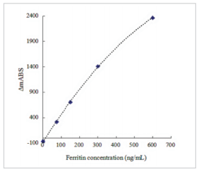
Fig. 2 Calibration curves for ferritinbyLETIA
Clinical Comparison
The CUSAg CLIA Ferritin assay is designed to have a correlation coefficient (r) of >0.90. A study was performed where lithium heparin plasma specimens were tested in replicates of three using different antibody combinations on the CUSAg CLIA platform and compared to a commercially available diagnostic kit (Comparison Assay). Data from this study were analyzed using the Passing-Bablok regression method and are summarized in the following table and scatted plot (Fig.3). Results show good parallelism between the two systems.
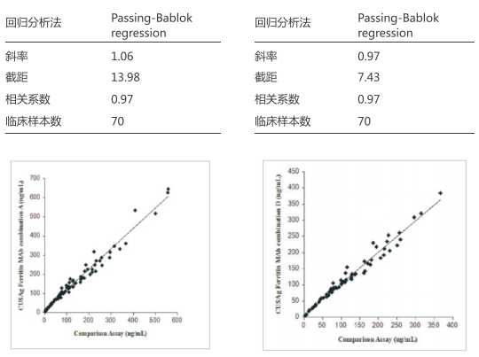
Fig.3Clinicalcomparison of CUSAg CLIA Ferritin assay andcommercial diagnosticassays
In order to meet the application of CUSAg anti-ferritin MAbs on LETIA platform, 35clinical blood samples were separately tested usingmixedMAbs on the CUSAgLETIAplatform and compared to a commercial diagnostic kit. Thecorrelation coefficient between the two systems is over 0.98.
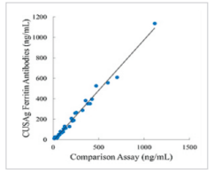
Fig.4 Comparison ofCUSAgLETIA ferritin assay andcommercial diagnosticassays
Thermal stability
Our anti-human ferritin monoclonal antibodies presented in PBS buffer without anypreservative were stored at -20°C, 2-8°C and 37°C for 15 days, respectively. During thisperiod,
the titers of four MAbs weredetermined, respectively. Fig.5 shows the relative titers of the MAbs treated at 37°C or 2-8°C were similar with that at-20°C. All the MAbs werestable from-20°C to 37°C.
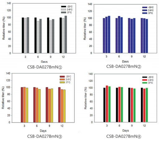
Fig.5 Evaluation ofanti-ferritin MAbs stability at different temperatures
Recombinant Ferritin Light Chain
Ferritin light chain is a protein that in humans is encoded by the FTLgene. This gene encodes the light subunit of the ferritin protein, Ferritin is the major intracellular iron protein in prokaryotes and eukaryotes. It is composed of 24 subunits of the heavy and light ferritin chains.The recombinant ferritin light chain protein (Cat:CSB-DP027B) is also offered by CUSAg. It could be used as calibrator in immunoassay and applied to western blotting.
References
1. Theil EC. Ferritin: structure, gene regulation, and cellular function in animals, plants, and microorganisms. Annual review of biochemistry.1987 56 (1): 289–315.
2. Guyatt GH, Patterson C, Turpie I et al. (1990). Diagnosis of iron-deficiency anemia in the elderly. Am J Med. 1990 88 (3): 205–9.doi:10.1016/0002-9343(90)90143-2.
3. Pongstaporn W, Bunyaratavej A.Hematological parameters, ferritin and vitamin B12 in vegetarians. J Med Assoc Thai. 1999 March 82 (3): 304–11.